Rotator Cuff Injuries
William T. Pennington, M.D.
The Orthopedic Institute of Wisconsin
What is the Rotator Cuff?
The rotator cuff is comprised of the tendons of four muscles that originate on the scapula (shoulder blade) that attach to the proximal humerus. The functioning rotator cuff will keep the humeral head (ball) centered in relation to the glenoid surface (cup) during shoulder motion while also serving to provide muscular force in planes of shoulder motion including forward flexion, abduction, internal and external rotation.
The muscles that make up the rotator cuff are as follows:
Supraspinatus muscle: Initiates shoulder flexion and abduction, most commonly torn tendon.
Infraspinatus muscle: External Rotator of the shoulder.
Teres Minor muscle: External rotator of the shoulder.
Subscapularis muscle: Internal rotator of the shoulder.
What happens when the rotator cuff is injured?
Rotator cuff injuries that are typically encountered are those that involve inflammation of the tendon (tendinopathy) and overlying bursa (bursitis) or tearing of the rotator cuff attachment to the proximal humerus. Patients often present with a painful shoulder that is worsened with any type of reaching or
lifting of the arm. Typically patients will complain of pain on the top of the shoulder with radiation of pain down the front and side of the arm in the biceps
and deltoid region. When the tendinous attachment of the rotator cuff is torn away from the proximal humerus this pain can be accompanied by significant weakness during arm motion or even the inability to move the arm actively is certain planes.
Inflammation of the rotator cuff can occur from episodes of increased activities resulting in an acutely painful state or inflammation can result from prolonged insults to the shoulder that occur during everyday activities. An extrinsic factor that can predispose patients to rotator cuff injuries and inflammation is the presence of a “bone spur” on the undersurface of the acromion, the bone directly over the rotator cuff, which can cause mechanical impingement to the underlying rotator cuff during certain motions of the shoulder. This impingement can cause repeated injuries to the underlying rotator cuff tendons resulting in inflammation, tendinopathy, bursitis and sometimes tears of the rotatorcuff.
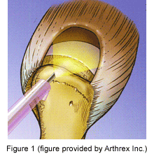 Tears of the rotator cuff typically occur in varying degrees of magnitude. They can be partial thickness in nature in which a portion of the thickness of the tendinous attachment is torn away from the bony attachment or full thickness in which the entire thickness of the rotator cuff tendon is torn away from the proximal humerus. Tears can involve only a portion of a single tendon or they can be so large that they can involve the entire tendinous attachment of one or more of the rotator cuff tendons (Figure 1). The attachment site of the rotator cuff tendon to bone is an area that does not have any blood supply, therefore, rotator cuff tears don’t typically heal spontaneously back to the bone.
How do we treat rotator cuff problems?
It is difficult to have a set treatment protocol for all rotator cuff injuries as each case needs to be considered on an individual basis to determine the most appropriate course of action that should be employed. When presented with apatient complaining of shoulder pain that appears to be attributable to the rotator cuff region we need to collect data that will help us diagnose the problem and hopefully prescribe a regimen that will eventually result in the
patient regaining lost function and experience pain relief.
We typically begin with a patient history and physical examination focusing on when the problem occurred, how it occurred, what makes it better or worse and what has been attempted up to the point of our evaluation to try to make things better. We examine the patient focusing on appearance, range of motion both active and passive, strength of the shoulder in all of the motion planes, palpation of the rotator cuff attachment, and finally we perform certain physical examination tests that help predictably diagnose the problem. We will often obtain standard radiographs (x-rays) of the shoulders to evaluate for the presence of the spurs
on the undersurface of the acromion and to also evaluate for any other bony problems such as arthritis that may be contributing to the painful condition. It
is very important to also evaluate the cervical spine (neck) region to also rule out any problem in that area that may be causing the current pain in the shoulder. Occasionally, if an acute traumatic event such as a fall precipitated the pain and we’re concerned that this traumatic event caused a rotator cuff
tear in a young active patient we’ll proceed to ordering advanced imaging techniques such as a MRI scan to thoroughly evaluate the rotator cuff prior to instituting a treatment plan.
Treatment of rotator cuff injuries often begins with a period of rest from activities that may exacerbate the pain. We often utilize anti-inflammatory pills if possible to help with the pain and inflammation and prescribe physical therapy. The physical therapist will focus on re-gaining or maintaining motion of the shoulder joint that may diminish due to the painful state of the affected shoulder. They often also employ modalities such as ultrasound or iontophoresis to help with the inflamed state providing pain relief. When the patient has a shoulder that has a full range of motion and has reasonable pain relief the therapist will then focus on strengthening the rotator cuff muscles as well as other muscles surrounding the region that may eventually lead to the restoration of function and proper kinematics of the shoulder joint hopefully leading to a complete functional recovery that is satisfactory to the patient. When a patient benefits from this type of treatment, typically this benefit begins to occur after four to six weeks of physical therapy. Sometimes the shoulder is too painful to tolerate the therapy and in these instances we may also suggest a cortisone injection to help with the pain so the patient is able to tolerate the pain while hopefully recovering without needing more aggressive treatment.
Cortisone injection is a commonly used, often feared, treatment that can be quite beneficial to assist patients in recovering from a rotator cuff injury or inflamed state. Cortisone is an injectable anti-inflammatory agent that we are able to administer directly to the site of injury hopefully resulting in significant pain relief and reduction in inflammation. We typically employ one injection in conjunction with other treatment modalities such as therapy to hopefully help the patient recover the function that they’re after. If another injection is necessary we will offer it twelve weeks after the initial injection only if the initial injection was beneficial and has worn off. We do not think that any more injections than two are beneficial due to possible weakening of
the rotator cuff with repeated injections (greater than three) and the significantly higher failure rate of surgeries that occurs in patients that eventually undergo rotator cuff repair surgery that have had more than three cortisone injections in the operative shoulder.
If all of the non-surgical treatment modalities fail, we do sometimes need to employ surgical methods to help facilitate recovery and restoration of shoulder function in patients afflicted with rotator cuff problems. Of note, there are some instances such as the young active patient with an acute traumatic full thickness rotator cuff tear that we typically recommend proceeding with surgical repair immediately. These patients tend to demonstrate better clinical outcomes with acute repair of these injuries rather than in the setting of delayed repair.
When considering rotator cuff repair an imaging study such as a MRI scan is useful to determine reparability of the rotator cuff by evaluating the retraction, tissue quality and associated atrophy of the muscle. This data is useful when preoperatively counseling the patient about perceived benefits of proceeding with an operative approach. As one would intuitively infer, those with larger retracted tears with associated muscle atrophy are less predictably repaired than those with smaller non-retracted tears with minimal to no muscle atrophy.
How do we surgically approach rotator cuff problems?
Surgical intervention in the instance of rotator cuff injuries is typically taken care of arthroscopically. Arthroscopy is a surgical approach utilizing a small camera, an arthroscope, to completely examine the shoulder structures including the rotator cuff to determine exactly what needs to be done. If there is a spur on the undersurface of the acromion causing impingement of the rotator cuff it is removed using a burr to alleviate the impingement and pain that the spur may be responsible for causing. The rotator cuff is thoroughly evaluated and if there is a tear the rotator cuff is repaired as well. Techniques have evolved that now allow us to repair most rotator cuff tears through an arthroscopic approach. By utilizing this technique of rotator cuff repair the advantage for patients is that there is less invasion of the overlying soft tissue, therefore, less trauma occurs and theoretically there is less of a chance that stiffness or loss of motion will occur post- operatively.
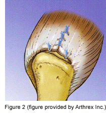
The technique of arthroscopic rotator cuff repair is depicted in Figure 2. Initially the arthroscope is inserted into the shoulder joint through a small incision in the back of the shoulder to assess the entire shoulder joint. There are instances that other injuries do exist that wouldn’t necessarily be seen if an open surgery was performed. If problems are seen with the arthroscope they can typically be addressed during the arthroscopy as well by placing instruments into the shoulder joint through a cannula placed through an anterior portal. After all of the pathology is addressed the rotator cuff tear is evaluated and the repair technique that will be employed is determined based on the anatomy of the tear. If there is a spur present a subacromial decompression is performed to alleviate any impingement present.
The rotator cuff tendon is generally repaired by reattaching the torn tendon back to the proximal humerus to the site that it was torn away from. The site of reattachment is referred to as the “footprint” of the rotator cuff. The footprint is the area on the humerus that the rotator cuff normally attaches to. When performing these repairs arthroscopically a device called a rotator cuff anchor is typically used to perform this reattachment. The anchor that we utilize is a screw made out of bioabsorbable plastic the we can insert into the proximal humerus just to the outside of the rotator cuff footprint. Prior to the insertion of this anchor a burr is used to remove the bone on the humerus that is overlying the footprint to create a trough in the footprint that has a bleeding surface. The anchor is then placed just outside this trough and sutures that are attached to the anchor are passed through the rotator cuff where it is torn away from the bone. After this process is complete the sutures are tied and this results in the reattachment of the rotator cuff tendon to the proximal humerus over the bleeding footprint.
 The repair technique is strong, however, it is not strong enough to withstand active use of the shoulder muscles that have been reattached. This process typically takes six weeks for the initial phases of healing to occur and at least another six weeks for the healing tissue to mature. The actual healing that takes place is that the bone on the footprint that is burred bleeds under the repaired rotator cuff resulting in a hematoma being formed that eventually leads to the healing of the rotator cuff to the footprint over time. Due to this healing and maturation process the repair needs to be protected so that the repair is not ruined prior to the time that healing occurs. Therefore, active forward elevation and sideways elevation actively is not allowed for 6 weeks post- operatively. We do allow passive motion of the shoulder and in fact utilize a machine that will provide this passive motion to help decrease the occurrence of stiffness that may occur in these patients post- operatively.
What is the post-operative protocol?
Patients are typically kept in a sling after surgery to serve as a reminder about the fact that they are not allowed to actively move the arm. Immediately post-operatively a polar ice pack is placed on the shoulder to help decrease the inflammation and pain. The continuous passive motion (CPM) machine that is supplied is generally started the day after surgery and advanced as the patients tolerate. The patient returns to the office five to seven days post-operatively for suture removal from their four small incisions and then return at the five week point for a passive motion check. Physical therapy typically begins three weeks after surgery and focuses on pain relief and passive motion initially followed by the advancement to active motion and strengthening of the repaired cuff when appropriate.
We have studied long term outcomes of arthroscopic rotator cuff repairs in patients and, if everything goes well, full recovery occurs in three to four months on average. Occasionally conditions such as excessive postoperative stiffness may prolong this recovery period.
In rare occasions the tear is so large that it is not reparable. In these instances sometimes debridement of the shoulder joint and rotator cuff tear with removal of spurs that may be present results in decreased pain in the shoulder. If severe arthritis is present with one of these massive irreparable tears a shoulder replacement procedure is sometimes required to provide pain relief that is desired.
Our goal at The Orthopedic Institute of Wisconsin is to provide high quality care both non-surgical and surgical that eventually will allow patients to regain lost function and experience pain relief that will hopefully result in the improvement of their quality of life. Through state of the art care our aim is to facilitate our patients return to a pre-injury level of function that ultimately will be satisfactory to our patients.
Disclaimer
This web site contains general medical information and does not replace the medical advice of your physician. If you have questions about your medical condition or exercises, ask your doctor or health care provider.